
Lightweight Pneumonia Detection Using Classical Descriptors on Edge AI Devices
DOI:
https://doi.org/10.33411/IJIST/1817Keywords:
Pneumonia detection, chest X-ray, handcrafted features., edge AI, machine learning, HOG, SIFT, ORB, Random Forest, SVM, Logistic RegressionAbstract
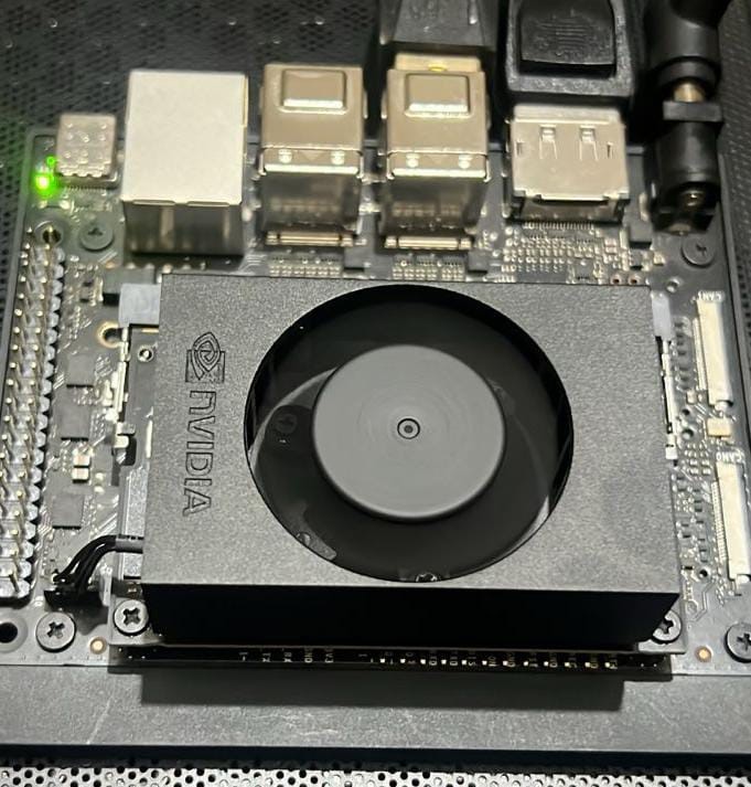
Pneumonia remains a pressing global health issue, and timely chest X-ray interpretation is central to effective patient care. This paper describes a lightweight pipeline for detecting pneumonia in chest radiographs using classical machine learning classifiers paired with handcrafted image features, aimed at hardware with tight processing budgets. Three widely used descriptors — HOG, SIFT, and ORB — were applied to the publicly available Kaggle chest X-ray dataset containing 5,863 labeled pediatric radiographs (4,273 Pneumonia, 1,590 Normal), pre-split into training (5,216), validation (16), and test (624) partitions. Four classifiers — Naive Bayes, SVM, Random Forest, and Logistic Regression — were trained with class-weight balancing to account for the roughly 2.7:1 class imbalance. Training and initial evaluation were carried out in Google Colab GPU-accelerated environment on a fixed held-out test set (approximately 89:11 train-to-test ratio), with 5-fold cross-validation on training data used to assess generalization. The strongest pipeline was subsequently ported to an NVIDIA Jetson Orin Nano Super for edge inference profiling. Random Forest with HOG features achieved the highest accuracy at 93.52% and an F1 score of 95.60%, while Logistic Regression with ORB features achieved the fastest inference speed at 0.187 ms per image (5,342.79 FPS). McNemar's test confirmed statistically significant gaps between the best and worst configurations (p < 0.01). Overall, the findings indicate that handcrafted features and traditional classifiers can produce clinically relevant performance within the power and memory envelopes of affordable edge hardware — a promising direction for pneumonia screening where specialist radiology support is scarce.
References
“Pneumonia - Our World in Data.” Accessed: Mar. 25, 2026. [Online]. Available: https://ourworldindata.org/pneumonia
Daniele Piovani, Gisella Figlioli, “The global burden of enteric fever, 2017–2021: a systematic analysis from the global burden of disease study 2021,” EClinicalMedicine, vol. 77, p. 102883, 2024, [Online]. Available: https://www.thelancet.com/journals/eclinm/article/PIIS2589-5370(24)00462-0/fulltext
“Pneumonia: Causes, Symptoms, Diagnosis & Treatment.” Accessed: Mar. 25, 2026. [Online]. Available: https://my.clevelandclinic.org/health/diseases/4471-pneumonia
A. D. Nguyen, D. R. Stamm, and H. A. Stankewicz, “Atypical Bacterial Pneumonia,” StatPearls, Apr. 2025, Accessed: Mar. 25, 2026. [Online]. Available: https://www.ncbi.nlm.nih.gov/books/NBK532239/
Pedro Cruz, Ana M Meireles, “COVID-19-Associated Cognitive Biases on Pneumonia Differential Diagnosis,” Cureus, vol. 16, no. 2, 2024, [Online]. Available: https://pubmed.ncbi.nlm.nih.gov/38558668/
“An Avoidable Cognitive Error in Chest Radiography - PMC.” Accessed: May 05, 2026. [Online]. Available: https://pmc.ncbi.nlm.nih.gov/articles/PMC10756156/
“Comparison of Chest Radiograph Impressions for Diagnosing Pneumonia: Accounting for Categories of Language Certainty - PubMed.” Accessed: May 05, 2026. [Online]. Available: https://pubmed.ncbi.nlm.nih.gov/35792164/
Cihan Aydin, Hafize Kızılkaya, “AI Models for Accurate Bacterial Pneumonia Diagnosis in Chest X-ray Images,” Bozok Tıp Derg., vol. 15, no. 2, pp. 169–177, 2025, doi: 10.16919/bozoktip.1593097.
Amer Kareem, Haiming Liu & Paul Sant, “Review on Pneumonia Image Detection: A Machine Learning Approach,” Human-Centric Intell. Syst., vol. 2, pp. 31–43, 2022, [Online]. Available: https://link.springer.com/article/10.1007/s44230-022-00002-2
Apurv Verma, D R Suman Kumar Swarnkar, “Advanced Deep Learning Models for Pneumonia Detection,” Proc. Int. Conf. Smart Heal. Intell. Technol., 2025, [Online]. Available: https://www.atlantis-press.com/proceedings/icshit-24/126010389#:~:text=To address these challenges%2C deep,in chest X-ray pictures.
S. Sharma and K. Guleria, “A systematic literature review on deep learning approaches for pneumonia detection using chest X-ray images,” Multimed. Tools Appl. 2023 838, vol. 83, no. 8, pp. 24101–24151, Aug. 2023, doi: 10.1007/s11042-023-16419-1.
S. Parveen and K. B. Khan, “Detection and classification of pneumonia in chest X-ray images by supervised learning,” Proc. - 2020 23rd IEEE Int. Multi-Topic Conf. INMIC 2020, Nov. 2020, doi: 10.1109/INMIC50486.2020.9318118.
T. B. Chandra and K. Verma, “Pneumonia Detection on Chest X-Ray Using Machine Learning Paradigm,” Adv. Intell. Syst. Comput., vol. 1022 AISC, pp. 21–33, 2020, doi: 10.1007/978-981-32-9088-4_3.
“Machine Learning Model Based on Radiomic Features for Differentiation between COVID-19 and Pneumonia on Chest X-ray - PubMed.” Accessed: May 05, 2026. [Online]. Available: https://pubmed.ncbi.nlm.nih.gov/36081170/
“In Search of an Efficient and Reliable Deep Learning Model for Identification of COVID-19 Infection from Chest X-ray Images - PubMed.” Accessed: May 05, 2026. [Online]. Available: https://pubmed.ncbi.nlm.nih.gov/36766679/
S. Hu, “Weakly Supervised Deep Learning for COVID-19 Infection Detection and Classification From CT Images,” IEEE Access, vol. 8, pp. 118869–118883, 2020, doi: 10.1109/ACCESS.2020.3005510.
A T ; Nagi, M J ; Awan, “Performance Analysis for COVID-19 Diagnosis Using Custom and State-of-the-Art Deep Learning Models,” Appl. Sci., vol. 12, no. 13, p. 6364, 2022, doi: https://doi.org/10.3390/app12136364.
Janmenjoy Nayak, Bighnaraj Naik, “Significance of deep learning for Covid-19: state-of-the-art review,” Res. Biomed. Eng., vol. 38, no. 1, pp. 243–266, 2021, doi: 10.1007/s42600-021-00135-6.
Neil C. Thompson, Kristjan Greenewald, Keeheon Lee, Gabriel F. Manso, “The Computational Limits of Deep Learning,” arXiv:2007.05558, 2020, [Online]. Available: https://arxiv.org/abs/2007.05558
T. Dhar, N. Dey, S. Borra, and R. Simon Sherratt, “Challenges of Deep Learning in Medical Image Analysis - Improving Explainability and Trust,” IEEE Trans. Technol. Soc., vol. 4, no. 1, pp. 68–75, Mar. 2023, doi: 10.1109/TTS.2023.3234203.
“Chest X-Ray Images (Pneumonia).” Accessed: Mar. 25, 2026. [Online]. Available: https://www.kaggle.com/datasets/paultimothymooney/chest-xray-pneumonia
N. Dalal and B. Triggs, “Histograms of oriented gradients for human detection,” Proc. - 2005 IEEE Comput. Soc. Conf. Comput. Vis. Pattern Recognition, CVPR 2005, vol. I, pp. 886–893, 2005, doi: 10.1109/CVPR.2005.177.
D. G. Lowe, “Distinctive image features from scale-invariant keypoints,” Int. J. Comput. Vis., vol. 60, no. 2, pp. 91–110, Nov. 2004, doi: 10.1023/B:VISI.0000029664.99615.94.
E. Rublee, V. Rabaud, K. Konolige, and G. Bradski, “ORB: An efficient alternative to SIFT or SURF,” Proc. IEEE Int. Conf. Comput. Vis., pp. 2564–2571, 2011, doi: 10.1109/ICCV.2011.6126544.
Corinna Cortes & Vladimir Vapnik, “Support-vector networks,” Mach. Learn., vol. 20, pp. 273–297, 1995, doi: https://doi.org/10.1007/BF00994018.
L. Breiman, “Random forests,” Mach. Learn., vol. 45, no. 1, pp. 5–32, Oct. 2001, doi: 10.1023/A:1010933404324/METRICS.
M. E. H. Chowdhury, “Can AI help in screening viral and COVID-19 pneumonia?,” IEEE Access, vol. 8, pp. 132665–132676, 2020, doi: 10.1109/ACCESS.2020.3010287.
Downloads
Published
How to Cite
Issue
Section
License
Copyright (c) 2026 50sea

This work is licensed under a Creative Commons Attribution 4.0 International License.