Preserving Spatial and Hierarchical Lesion Structures in Automated Skin Cancer Diagnosis through DermaCap and CCSA
DOI:
https://doi.org/10.33411/IJIST/1865Keywords:
Skin Lesion Classification, Automated Skin Cancer Diagnosis, Deep Learning in Dermatology, Hierarchical Lesion Feature Preservation, DermaCap Augmentation FrameworkAbstract
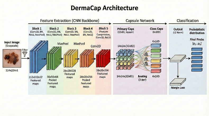
Early and reliable detection of skin cancer remains a global clinical priority, particularly for rare lesion categories that are often underrepresented due to limited data and severe class imbalance. To address these challenges and the limitations of traditional Convolutional Neural Networks (CNNs) in preserving spatial relationships, this study proposes DermaCap, a hybrid CNN–Capsule Network architecture designed to capture lesion pose, orientation, and hierarchical structural features. The framework follows a rare-class-first design philosophy, optimizing the model architecture, dataset curation, and training strategy to improve sensitivity toward diagnostically challenging lesions. Additionally, Clinically Constrained Structural Augmentation (CCSA) is introduced to enhance dataset diversity while preserving dermatologically meaningful morphology and color realism. The proposed model was evaluated using a carefully selected six-class subset of the HAM10000 dermatology database, which contains a total of 3,310 dermatoscopic images representing multiple diagnostic categories. The evaluation resulted in an approximate accuracy of 91%. The precision, recall, and F1-score show consistent values across both majority and minority classes. Therefore, the proposed model shows stable and consistent diagnostic performance for dermatoscopic image classification. Overall, DermaCap presents a clinically aligned, structure-aware approach for more trustworthy and scalable AI-assisted skin lesion diagnosis.
References
A. Lomas, J. Leonardi-Bee, and F. Bath-Hextall, “A systematic review of worldwide incidence of nonmelanoma skin cancer,” Br. J. Dermatol., vol. 166, no. 5, pp. 1069–1080, May 2012, doi: 10.1111/J.1365-2133.2012.10830.X.
J. A. Siegel RL, Miller KD, Fuchs HE, “Cancer statistics,” Cancer J Clin, vol. 72, no. 1, 2022, [Online]. Available: https://www.cancer.gov/about-cancer/understanding/statistics
“Analysis of trends in US dermatologist density and geographic distribution - PubMed.” Accessed: May 09, 2026. [Online]. Available: https://pubmed.ncbi.nlm.nih.gov/38574771/
A. Esteva et al., “Dermatologist-level classification of skin cancer with deep neural networks,” Nat. 2017 5427639, vol. 542, no. 7639, pp. 115–118, Jan. 2017, doi: 10.1038/nature21056.
S. Sabour, N. Frosst, and G. E. Hinton, “Dynamic Routing Between Capsules,” Nov. 2017, Accessed: May 09, 2026. [Online]. Available: http://arxiv.org/abs/1710.09829
“Skin cancer detection using deep machine learning techniques - ScienceDirect.” Accessed: May 09, 2026. [Online]. Available: https://www.sciencedirect.com/science/article/pii/S2666521224000589
M. M. Haque, R. Akter, A. S. M. A. S. Akib, and A. Hasib, “A Deep Learning Approach for Automated Skin Lesion Diagnosis with Explainable AI,” Jan. 2026, Accessed: May 09, 2026. [Online]. Available: http://arxiv.org/abs/2601.00964
D. C. Malo, M. M. Rahman, J. Mahbub, and M. M. Khan, “Skin Cancer Detection using Convolutional Neural Network,” 2022 IEEE 12th Annu. Comput. Commun. Work. Conf. CCWC 2022, pp. 169–176, 2022, doi: 10.1109/CCWC54503.2022.9720751.
F. Setiawan and B. Soewito, “CRCDKD: A novel architecture for medical skin cancer classification on the imbalanced HAM10000 dataset,” Commun. Math. Biol. Neurosci., vol. 2025, no., p. Article ID 93, 2025, doi: 10.28919/CMBN/9417.
S. Karimzadhagh et al., “Performance of Artificial Intelligence in Skin Cancer Detection: An Umbrella Review of Systematic Reviews and Meta-Analyses,” Int. J. Dermatol., vol. 65, no. 1, pp. 69–85, Jan. 2026, doi: 10.1111/IJD.17981.
“Leveraging machine learning to predict de novo skin malignancy following lung transplantation - PubMed.” Accessed: May 09, 2026. [Online]. Available: https://pubmed.ncbi.nlm.nih.gov/41553243/
A. Gupta, K. Bansal, Arti, A. Sabharwal, S. R. N. Reddy, and R. Anand, “Advancing melanoma detection with transfer learning and hybrid models,” Netw. Model. Anal. Heal. Informatics Bioinforma. 2025 151, vol. 15, no. 1, pp. 8-, Dec. 2025, doi: 10.1007/S13721-025-00683-2.
“Skin Cancer MNIST: HAM10000.” Accessed: May 09, 2026. [Online]. Available: https://www.kaggle.com/datasets/kmader/skin-cancer-mnist-ham10000
Downloads
Published
How to Cite
Issue
Section
License
Copyright (c) 2026 50sea

This work is licensed under a Creative Commons Attribution 4.0 International License.