ScaleCeph: A Two-Stage Coarse-to-Fine ROI Cascade for Cross-Device Cephalometric Landmark Localization
Keywords:
Automated Cephalometric Analysis, Cephalometric Landmark Localization, Cross-Device Robustness, Deep Learning, HRNetAbstract
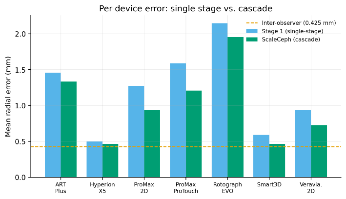
Cephalometric landmark localization is an important part of orthodontic diagnosis and treatment planning, but manual annotation is slow and has notable inter-observer variation. Automated methods have advanced rapidly, yet most are developed and tested on images from a single device and report a single aggregate error, leaving open how they behave when a clinic pools radiographs from several machines with different fields of view, contrast, and pixel spacing. We present ScaleCeph, a two-stage coarse-to-fine cascade that treats this cross-device variation as the central problem. A first HRNet-W32 localizes 19 landmarks on the whole radiograph; the bounding box of these predictions defines a craniofacial region of interest that is cropped and magnified to a canonical scale, absorbing the inter-device differences, and a second, independently trained network refines the landmarks on this magnified view. Coordinates are decoded with a robust windowed soft-argmax decoder, and ablations show that the design choices, prediction-driven crops, sharp σ = 2 heatmap targets with a weighted-BCE loss, and the windowed decoder, are each necessary rather than incidental. On the Aariz benchmark (1,000 cephalograms, seven devices), ScaleCeph attains a mean radial error (MRE) of 1.076 ± 1.18 mm (95% CI 0.99–1.16 mm) and 88.8% successful detection rate (SDR) within 2 mm (95% CI 87.7–90.0), against the single-stage baseline (1.252 ± 1.17 mm, 85.1%; paired Wilcoxon signed-rank p < 10⁻²⁰), and raises the macro-averaged per-device SDR@2 mm from 85.5% to 89.5%. On the best-resolved machines performance approaches inter-observer agreement, supporting prediction-driven scale normalization for robust cross-device localization.
References
“Does orthodontic treatment affect patients’ quality of life? - PubMed.” Accessed: Aug. 10, 2026. [Online]. Available: https://pubmed.ncbi.nlm.nih.gov/18676797/
J. Monisha, U. Sangeetha, B. Nivethitha, and B. Madhan, “Agreement between cephalometric analyses in diagnosing the dento-skeletal characteristics of malocclusion,” J. Oral Biol. Craniofacial Res., vol. 15, no. 4, pp. 744–748, Jul. 2025, doi: 10.1016/j.jobcr.2025.04.012.
H. Zhang et al., “Deep Learning Techniques for Automatic Lateral X-ray Cephalometric Landmark Detection: Is the Problem Solved?,” Sep. 2024, Accessed: Jul. 27, 2026. [Online]. Available: https://arxiv.org/pdf/2409.15834
Q. Chang, Z. Wang, F. Wang, J. Dou, Y. Zhang, and Y. Bai, “Automatic analysis of lateral cephalograms based on high-resolution net,” Am. J. Orthod. Dentofac. Orthop., vol. 163, no. 4, pp. 501-508.e4, Apr. 2023, doi: 10.1016/J.AJODO.2022.02.020.
M. Han et al., “Automated Landmark Detection and Lip Thickness Classification Using a Convolutional Neural Network in Lateral Cephalometric Radiographs,” Diagnostics 2025, Vol. 15, Page 1468, vol. 15, no. 12, p. 1468, Jun. 2025, doi: 10.3390/DIAGNOSTICS15121468.
M. S. I. Sumon et al., “Self-CephaloNet: a two-stage novel framework using operational neural network for cephalometric analysis,” Neural Comput. Appl. 2025 3716, vol. 37, no. 16, pp. 9777–9805, Mar. 2025, doi: 10.1007/S00521-025-11097-6.
Y. Shimamura et al., “Accuracy of cephalometric landmark and cephalometric analysis from lateral facial photograph by using CNN-based algorithm,” Sci. Reports 2024 141, vol. 14, no. 1, pp. 31089-, Dec. 2024, doi: 10.1038/s41598-024-82230-z.
S. H. Han et al., “Accuracy of posteroanterior cephalogram landmarks and measurements identification using a cascaded convolutional neural network algorithm: A multicenter study,” Korean J. Orthod., vol. 54, no. 1, pp. 48–58, 2024, doi: 10.4041/KJOD23.075.
S. Kang, I. Kim, Y. J. Kim, N. Kim, S. H. Baek, and S. J. Sung, “Accuracy and clinical validity of automated cephalometric analysis using convolutional neural networks,” Orthod. Craniofac. Res., vol. 27, no. 1, pp. 64–77, Feb. 2024, doi: 10.1111/OCR.12683.
R. Khan, M. A. Khalid, K. Zulfiqar, U. Bashir, and M. M. Fraz, “Enhancing Cephalometric Landmark Detection with a Two-Stage Cascaded CNN on Multi-resolution Multi-modal Data,” Lect. Notes Comput. Sci. (including Subser. Lect. Notes Artif. Intell. Lect. Notes Bioinformatics), vol. 14860 LNCS, pp. 3–18, 2024, doi: 10.1007/978-3-031-66958-3_1/SAVE-RESEARCH.
M. A. Khalid, A. Khurshid, K. Zulfiqar, U. Bashir, and M. M. Fraz, “A two-stage regression framework for automated cephalometric landmark detection incorporating semantically fused anatomical features and multi-head refinement loss,” Expert Syst. Appl., vol. 255, p. 124840, Dec. 2024, doi: 10.1016/J.ESWA.2024.124840.
S. Rashmi, S. Srinath, R. Rakshitha, and B. V. Poornima, “Ensemble learning methods with single and multi-model deep learning approaches for cephalometric landmark annotation,” Discov. Artif. Intell. 2024 41, vol. 4, no. 1, pp. 93-, Nov. 2024, doi: 10.1007/S44163-024-00207-3.
H. Zhu, Q. Yao, L. Xiao, and S. K. Zhou, “You Only Learn Once: Universal Anatomical Landmark Detection,” Lect. Notes Comput. Sci. (including Subser. Lect. Notes Artif. Intell. Lect. Notes Bioinformatics), vol. 12905 LNCS, pp. 85–95, Mar. 2022, doi: 10.1007/978-3-030-87240-3_9.
Z. Zhu et al., “An ensemble-based deep learning method through multi-scale cross-attention training for cephalometric landmark localization on lateral X-ray images,” Mar. 2025, doi: 10.21203/RS.3.RS-6105085/V1.
Z. Jiao et al., “Deep learning for automatic detection of cephalometric landmarks on lateral cephalometric radiographs using the Mask Region-based Convolutional Neural Network: a pilot study,” Oral Surg. Oral Med. Oral Pathol. Oral Radiol., vol. 137, no. 5, pp. 554–562, May 2024, doi: 10.1016/j.oooo.2024.02.003.
S. A. Prativi, A. B. Suksmono, T. L. E. Rajab, D. Danudirdjo, and A. Laviana, “Automatic landmark detection in cephalometric lateral radiograph using deep learning one-stage detectors,” 2024 14th Int. Conf. Syst. Eng. Technol. ICSET 2024 - Proceeding, pp. 67–72, 2024, doi: 10.1109/ICSET63729.2024.10775273.
I. Tafala, F. E. Ben-Bouazza, A. Edder, O. Manchadi, M. Et-Taoussi, and B. Jioudi, “Cephalometric Landmarks Identification Through an Object Detection-based Deep Learning Model,” Int. J. Adv. Comput. Sci. Appl., vol. 15, no. 2, pp. 859–867, 2024, doi: 10.14569/IJACSA.2024.0150286.
A. Jaheen et al., “CephRes-MHNet: A Multi-Head Residual Network for Accurate and Robust Cephalometric Landmark Detection,” Nov. 2025, Accessed: Jul. 27, 2026. [Online]. Available: https://arxiv.org/abs/2511.10173v2
F. Laitenberger, H. T. Scheuer, H. A. Scheuer, E. Lilienthal, S. You, and R. E. Friedrich, “Cephalometric landmark detection using vision transformers with direct coordinate prediction,” J. Cranio-Maxillofacial Surg., vol. 53, no. 9, pp. 1518–1529, Sep. 2025, doi: 10.1016/J.JCMS.2025.05.021.
H. Wu et al., “Cephalometric Landmark Detection across Ages with Prototypical Network,” Jun. 2024, Accessed: Jul. 27, 2026. [Online]. Available: https://arxiv.org/pdf/2406.12577
M. A. Khalid et al., “A Benchmark Dataset for Automatic Cephalometric Landmark Detection and CVM Stage Classification,” Sci. Data 2025 121, vol. 12, no. 1, pp. 1336-, Jul. 2025, doi: 10.1038/s41597-025-05542-3.
C. W. Wang et al., “A benchmark for comparison of dental radiography analysis algorithms,” Med. Image Anal., vol. 31, pp. 63–76, Jul. 2016, doi: 10.1016/J.MEDIA.2016.02.004.
J. Wang et al., “Deep High-Resolution Representation Learning for Visual Recognition,” IEEE Trans. Pattern Anal. Mach. Intell., vol. 43, no. 10, pp. 3349–3364, Aug. 2019, doi: 10.1109/TPAMI.2020.2983686.
Q. Wu, S. Y. Yeo, Y. Chen, and J. Liu, “Revisiting Cephalometric Landmark Detection from the view of Human Pose Estimation with Lightweight Super-Resolution Head,” Sep. 2023, Accessed: Aug. 10, 2026. [Online]. Available: https://arxiv.org/pdf/2309.17143
R. Chen, Y. Ma, N. Chen, D. Lee, and W. Wang, “Cephalometric Landmark Detection by AttentiveFeature Pyramid Fusion and Regression-Voting,” Lect. Notes Comput. Sci. (including Subser. Lect. Notes Artif. Intell. Lect. Notes Bioinformatics), vol. 11766 LNCS, pp. 873–881, Aug. 2019, doi: 10.1007/978-3-030-32248-9_97.
Downloads
Published
How to Cite
Issue
Section
License
Copyright (c) 2026 50sea

This work is licensed under a Creative Commons Attribution 4.0 International License.